Which microscope is more suitable for you? TEM or SEM
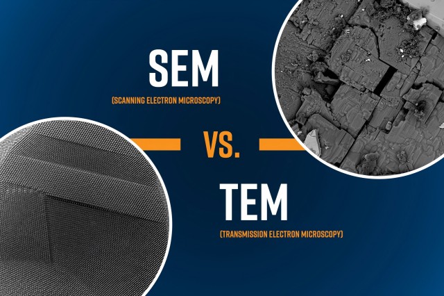
Transmission Electron Microscopes (TEM) and Scanning Electron Microscopes (SEM) are indispensable tools in modern scientific research. Compared to optical microscopes, electron microscopes offer higher resolution, allowing for the observation and study of specimens’ microstructure at a smaller scale.
Electron microscopes can provide high-resolution and high-magnification images by utilizing the interactions between an electron beam and a specimen. This enables researchers to obtain critical information that may be difficult to obtain through other methods.
Which microscope is more suitable for you?

When choosing the appropriate electron microscopy technique for your needs, various factors need to be considered to determine the best fit. Here are some considerations that can help you make a decision:

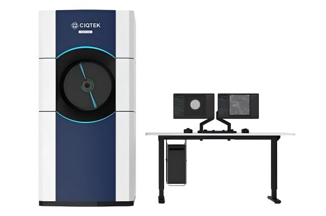
Field Emission TEM | TH-F120
Analysis Purpose:
First, it is important to determine your analysis purpose. Different electron microscopy techniques are suitable for different types of analysis.
a. If you are interested in surface features of a specimen, such as roughness or contamination detection, a Scanning Electron Microscope (SEM) may be more suitable.
b. However, a transmission electron microscope (TEM) may be more appropriate if you want to understand the crystal structure of a specimen or detect structural defects or impurities.
Resolution Requirements:
Depending on your analysis requirements, you may have specific resolution needs. In this regard, TEM generally has a higher resolution capability compared to SEM. If you need to perform high-resolution imaging, especially for observing fine structures, TEM may be more suitable.
Specimen Preparation:
An important consideration is the complexity of specimen preparation.
a. SEM specimens typically require minimal or no preparation, and SEM allows for more flexibility in specimen size, as they can be directly mounted on the specimen stage for imaging.
b. In contrast, the specimen preparation process for TEM is much more complex and requires experienced engineers to operate. TEM specimens must be extremely thin, typically below 150 nm, or even lower than 30 nm, and as flat as possible. This means that TEM specimen preparation may require more time and expertise.
Type of Images:

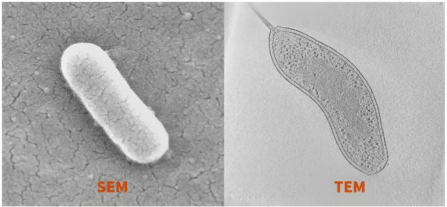
SEM provides detailed three-dimensional images of the specimen surface, while TEM provides two-dimensional projection images of the internal structure of the specimen.
a. Scanning Electron Microscope (SEM) provides three-dimensional images of the surface morphology of the specimen. It is mainly used for morphology analysis. If you need to examine the surface morphology of a material, SEM can be used, but you need to consider the resolution to see if it meets your experimental requirements.
b. If you need to understand the internal crystal or atomic structure of a material, TEM is required.
Transmission electron microscopy (TEM) is similar to a conventional microscope, providing two-dimensional images. It allows for observation of both the surface and inner layers of a specimen but lacks the three-dimensional aspect.
Difference:
Scanning Electron Microscope (SEM) observes the surface morphology of a specimen, while Transmission Electron Microscope (TEM) examines the structural morphology of a specimen.
Generally, TEM offers higher magnification and requires a higher vacuum. SEM can accommodate larger-sized specimens, with maximum diameters reaching over 200 mm and heights around 80 mm, whereas TEM specimens are typically placed on a grid with a diameter of around 3 mm for observation