Introduction to the Focused Ion Beam (FIB) Principle
Focused Ion Beam (FIB) is a microfabrication instrument that utilizes an electron lens to focus an ion beam into a very small size for precision cutting.
- Working Principle
Liquid Metal Ion Source
The ion source is the heart of the FIB system, and the accurate focusing of the ion beam begins with the emergence of liquid metal ion sources. The ions generated by liquid metal ion sources, mostly utilizing gallium (Ga) as the metal material, have high brightness and petite source sizes. Liquid metal ion sources are formed by the field-induced ion emission from a liquid metal surface under a strong electric field.
In the manufacturing process of the source, a tungsten wire with a diameter of about 0.5 mm is electrochemically etched to create a tungsten needle with a tip diameter of only 5-10 μm. The molten liquid metal is then adhered to the tip of the tungsten needle. Under the application of a strong electric field, the liquid metal forms a tiny tip (Taylor cone) due to the electric field force, with an electric field intensity of up to 10^10 V/m. Under such a high electric field, metal ions on the liquid surface evaporate into an ion beam through field evaporation. Despite the low ion current of only a few microamperes, the current density can reach approximately 106 A/cm2, and the brightness is about 20 μA/sr.
Focused Ion Beam System
Focused ion beam technology utilizes electrostatic lenses to focus the ion beam into a very small size for microfabrication. Commercial FIB systems typically extract the particle beam from liquid metal ion sources. Gallium (Ga) is often used as a metal material due to its low melting point, low vapor pressure, and good oxidation resistance.
By applying an external electric field (Suppressor) to the top of the ion column, a small tip of the liquid metal or alloy can be formed. With the negative electric field (Extractor) applied, the tip of the metal or alloy is pulled out to generate the ion beam. The ion beam is then focused using electrostatic lenses, and the size of the ion beam can be controlled by an Automatic Variable Aperture (AVA) with an adjustable aperture. The desired ion species are selected using an E×B mass analyzer. Finally, the ion beam is focused on the specimen and scanned using an octupole deflector and an objective lens. The ion beam bombards the specimen, and the secondary electrons and ions generated are collected, imaged, or used for physical sputtering, cutting, or grinding.
- Basic Functions
The basic functions of a focused ion beam microscope can be divided into four categories:
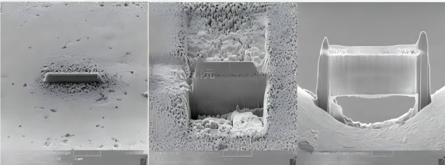
1. Precisional Cutting: Achieving cutting through the physical collision of ions. Widely used in Cross-Section processing and analysis of Integrated Circuits (ICs) and LCDs.
2. Selective Deposition: Decomposing organic metal vapor or gas-phase insulating material with the energy of the ion beam to locally deposit conductive or non-conductive materials. This process can provide deposition of metals and oxide layers, with common metals being platinum (Pt) and tungsten (W).
3. Enhanced Etching or Selective Etching: Accelerating cutting efficiency or selectively removing materials by using corrosive gases.
4. End Point Detection: Detecting the signals of secondary ions to understand the progress of cutting or etching.
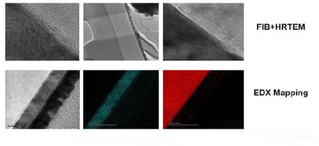
In novel focused ion beam microscopes, dual-beam models (ion beam + electron eam) have emerged. When using the ion beam for cutting, the electron beam can be used for imaging to not only prevent further “damage on-site” by the ion beam but also improve the image resolution effectively. They can also be equipped with X-ray spectrometers or secondary ion mass spectrometers for elemental analysis. The diverse analytical functions greatly enhance the convenience and utilization of focused ion beam microscopes.